How does an optical microscope work?
A simple microscope has one lens and is essentially a loupe or magnifying glass with a relatively high magnification.
A microscope is composed of four important parts - the light producing unit, which uses tungsten-halogen, or arc discharge. This light is collected by the condenser lens, which focuses the light onto the specimen. Presence of condensers makes the image much sharper.
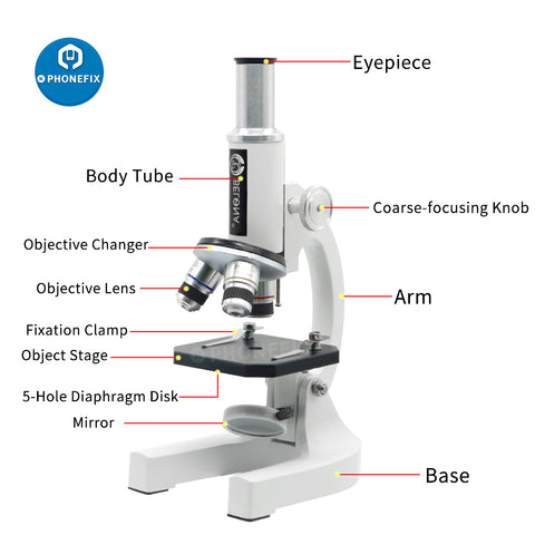
The basic modern microscope found in schools, hospitals, and research centers is a compound microscope which has a series of lenses to collect and focus the light transmitted through the specimen.

The optical or light microscope uses visible light transmitted through, refracted around, or reflected from a specimen.
The objective is the first lens that light from the spcimen passes through. It is the most complex to construct, and is made to have a short focal length. The final lens is the eye piece, which further magnifies the image, and creates an image perceivable by the human eye.

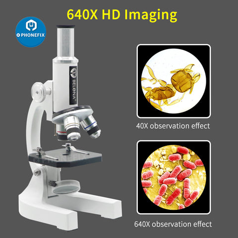
The aperture of a microscope determines its resolving power. Resolution is the ability of a microscope to keep all specific details of the object intact in the image. Hence, all microscopes, based on the technique being used, have a magnification limit at which there is no longer more resolution.
Simple ways to get around this is through techniques like oil immersion, which create a refractive index difference to bend the light inwards, and increase the magnification limit.
Light waves are chaotic; an incandescent light source emits light waves traveling in different paths and of varying wavelengths. Some of the lenses in a microscope bend these light waves into parallel paths, magnify and focus the light at the ocular.

Magnification is expressed in numeric multiples of how much enlargement occurs with a lens. If the magnification of a lens is 2X then it roughly doubles the size of the image of the object.
In many ways, an electron microscope functions similarly to an optical scope except that, instead of visible light, a stream of electrons is used to illuminate the specimen. The electron beam is focused with magnetic lenses. Changes to the electron beam inside the specimen are recorded and an image is formed based upon these changes.


Comments
Post a Comment